
LSIL — Low-Grade Squamous Intraepithelial Lesion
Also called mild dysplasia, or CIN 1 in its histologic equivalent. It is one of the most frequent cervical cytology results and, in the vast majority of cases, is related to a transient HPV infection that the body clears on its own.
Three fields from the same case.
Cervical cytology, Papanicolaou stain. Click the thumbnails to switch fields.
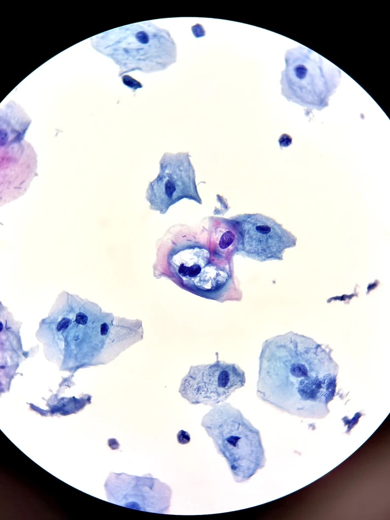
Field 1 — koilocytes: a cell with a well-defined perinuclear halo and an enlarged, hyperchromatic nucleus, the most characteristic cytologic finding of LSIL. Field 2 — binucleated cells (two nuclei in the same cell), another typical criterion, over mature cytoplasm. Field 3 — a superficial cell with mild nuclear atypia among unremarkable intermediate cells, illustrating the usually preserved background.
Your result, explained without fear.
Is it serious?
In the vast majority of cases, no. LSIL is a mild, very common cellular change. Most cases clear up on their own within one to two years, especially in younger people, with no treatment needed.
Why did this happen to me?
It is almost always caused by infection with the human papillomavirus (HPV). This is an extremely common infection — most sexually active people acquire it at some point in their lives, often without knowing it and without symptoms.
What happens now?
Your doctor or midwife will decide the most appropriate follow-up based on your age, history and current protocols — this could mean repeating the cytology in a few months, doing an HPV test, or referring you for colposcopy to look at the cervix more closely. There is no single right answer for every case.
Does this mean I have cancer?
No. LSIL is not cancer, nor an advanced precancerous lesion. It is the cytologic expression of an active HPV infection. The risk of progression to something more serious exists but is low, and that is exactly what medical follow-up monitors for.
Cytologic criteria and correlation.
Cytomorphologic criteria (Bethesda 2014): mature squamous cells (superficial/intermediate type) with nuclear enlargement of at least 3 times the nucleus of a normal intermediate cell; mild to moderate nuclear hyperchromasia; mild nuclear membrane irregularity; occasional binucleation or multinucleation; and perinuclear cytoplasmic cavitation with a well-defined, dense peripheral cytoplasmic rim (koilocytotic atypia) — the pathognomonic finding of the HPV cytopathic effect.
Histologic correlate: cervical intraepithelial neoplasia grade 1 (CIN 1) / mild dysplasia, when biopsied.
Differential diagnosis: reactive/reparative changes, atrophy (especially perimenopausal), and distinction from ASC-US when koilocytotic atypia is inconclusive — LSIL requires the characteristic nuclear criteria, not isolated perinuclear cavitation alone.
Natural history and follow-up: most LSIL regresses spontaneously, with higher regression rates in younger patients. Risk-based management (age, HPV co-testing, prior history) follows the guidelines in force in each country or society — for example ASCCP in the U.S., or local gynecology/pathology society recommendations — and does not reduce to a single universal algorithm.
HPV, in summary.
The cause behind the vast majority of LSIL cases, explained with what we actually know about it.
The human papillomavirus (HPV) is a group of more than 200 viral genotypes, of which around 40 infect the genital tract. It spreads mainly through sexual contact and is the most common sexually transmitted infection in the world: most sexually active people become infected at some point in their lives.
Genotypes are classified as low risk (associated with genital warts and mild cellular changes such as LSIL) and high risk (with oncogenic potential, associated with high-grade lesions and, if they persist unresolved for years, with cervical cancer). LSIL can appear with infection by either group — the cytologic finding reflects the virus's cytopathic effect on the cell, not directly its oncogenic risk.
Most HPV infections are transient: the immune system clears them within one to two years without leaving sequelae. Only when a high-risk genotype infection persists over a long period does the risk of progression to higher-grade lesions meaningfully increase. That is why clinical follow-up exists: to identify that minority of cases that do not resolve on their own.
HPV vaccination, ideally given before the start of sexual activity, very significantly reduces infection by the high-risk genotypes most frequently implicated in high-grade lesions and cervical cancer — it is one of the most effective primary prevention tools available in oncology.
What is the "Bethesda System"?
It is the standardized system used by laboratories around the world to report cervical cytology results, so that a result means the same thing at any center. It categorizes squamous findings into, among others: ASC-US, LSIL, ASC-H, HSIL and squamous carcinoma — each with defined cytologic criteria and an associated clinical management pathway. It is periodically updated; the current edition is from 2014.
Where this information comes from.
Recognized academic and clinical references, so you can verify it yourself.
- PathologyOutlines.com — cervical cytology section, diagnostic criteria for LSIL.pathologyoutlines.com
- Nayar R, Wilbur DC (eds.). The Bethesda System for Reporting Cervical Cytology: Definitions, Criteria, and Explanatory Notes, 3rd edition. Springer, 2015.
- ASCCP (American Society for Colposcopy and Cervical Pathology) — risk-based management guidelines for abnormal cytology results.asccp.org
- World Health Organization — information on HPV and cervical cancer prevention.who.int